Silicon becomes a landscape, crystals resemble foliage and fluorescent signals reveal the structures of cells. These selected images explore the meeting of scientific observation and visual imagination. Select an image to discover the material, technique and research behind it.
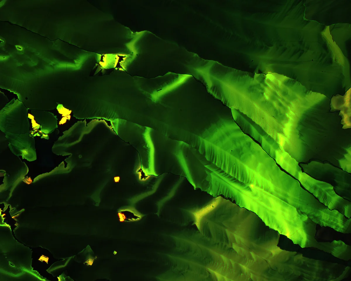
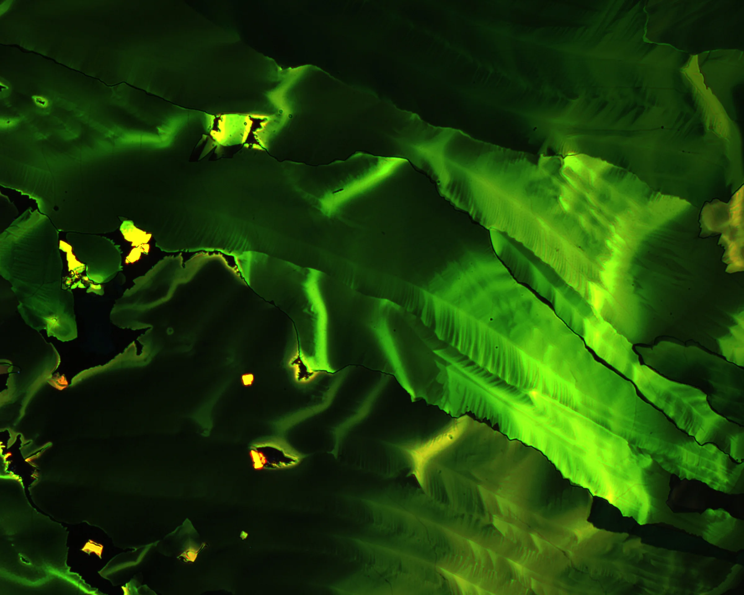
A hot solution of tetracene in toluene was cast onto glass. As the solution cooled and the solvent evaporated, crystalline films formed on the surface. Polarised-light microscopy reveals their varied patterns and textures.
Image: Qian Miao
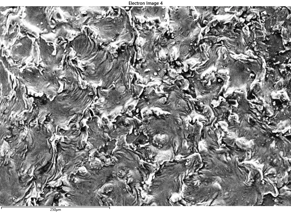
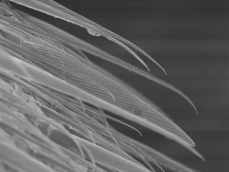
This is a silicon wafer after partial melting and recrystallisation. What looks like a disturbed water surface is a solid material with an intricate texture. The image draws attention to the patterns that remain when a material melts and forms crystals again.
Image: Ian Pong
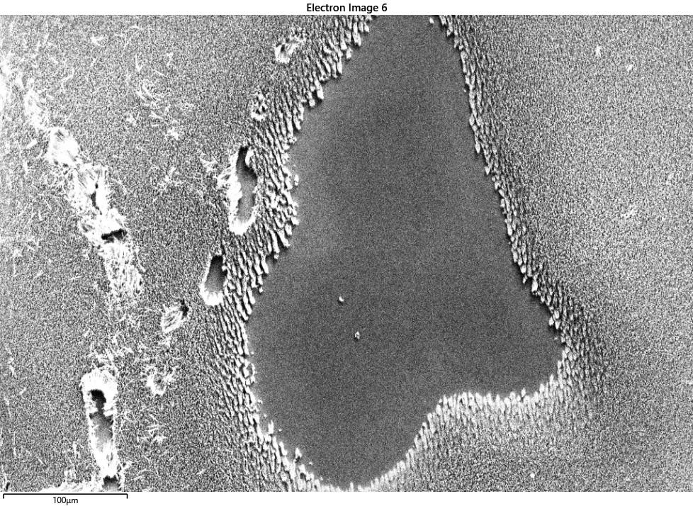
Carbon nanotubes have been grown on a silicon wafer. These tiny structures consist of carbon atoms arranged into tubes; grown together, they form a forest-like surface. The title invites a landscape reading of a specimen made on a very different scale.
Image: Ian Pong
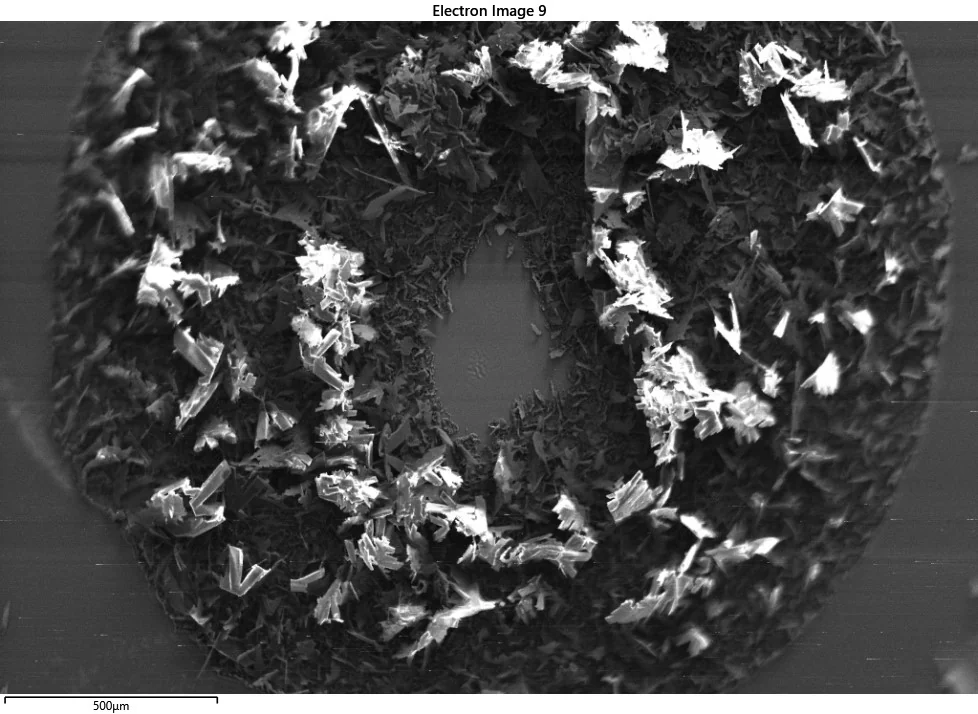
Carbon nanotubes grown on silicon form the structure in this image. The specimen was treated with SARS-CoV-2 spike protein for mass-spectrometric analysis. Its wreath-like appearance and title connect the material under investigation with remembrance of the pandemic.
Image: Ian Pong
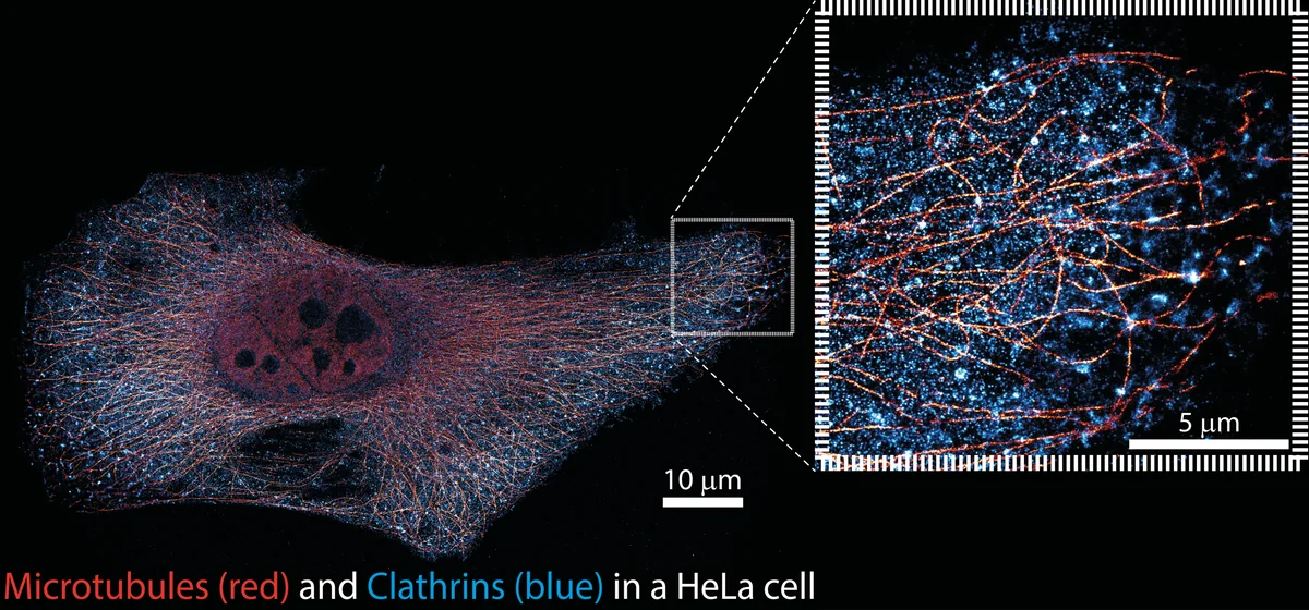
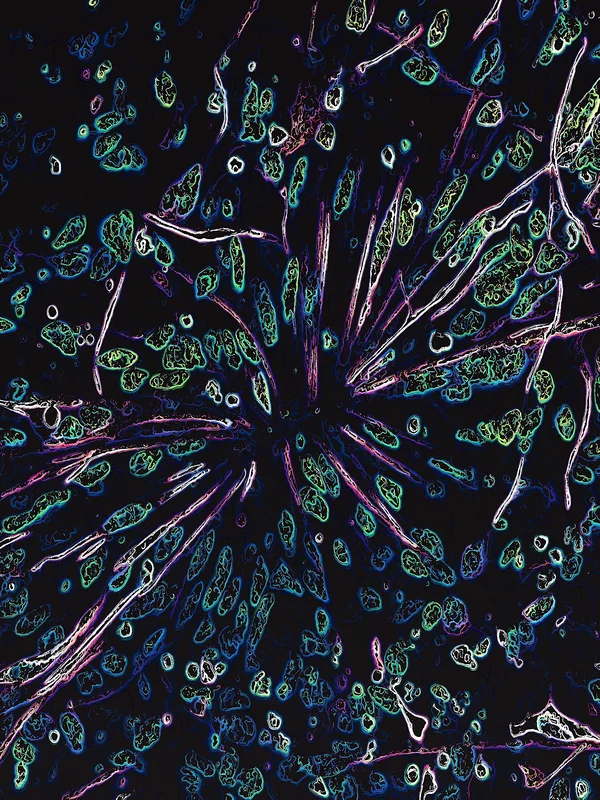
This image brings together multiple views of structures within human cells using exchange-PAINT, a super-resolution imaging technique. Combining the views allows different cellular components to be seen together at a level of detail beyond conventional light microscopy.
Image: Jeff Lam
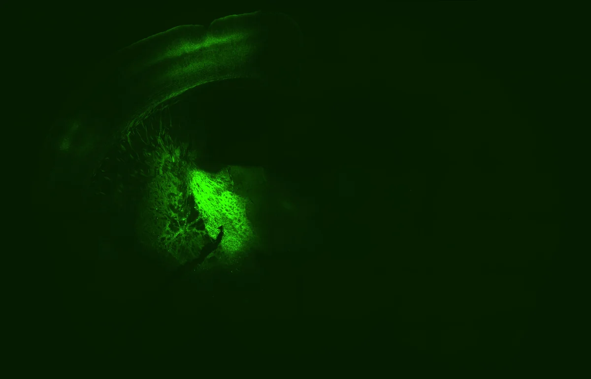
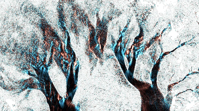
The image shows axonal projections from neurons in the globus pallidus. Viral delivery of channelrhodopsins tagged with a fluorophore makes these extensions visible. The branching paths reveal how a neuron reaches beyond its cell body.
Image: Yau Ming Chang
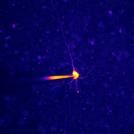
This image shows pyramidal neurons in layers five and six of a mouse’s prefrontal cortex, recorded using calcium imaging. The technique allows researchers to study changes in calcium within cells. The displayed colours should not, by themselves, be read as a calibrated measure of neuronal activity.
Image: Yau Ming Chang
Qian Miao is a professor of chemistry at the Chinese University of Hong Kong. He designs and makes organic molecules, exploring their structures and potential uses in electronic materials. A Croucher Senior Research Fellowship supported his research in 2019. His winning image, Crystalline Film of Tetracene, reveals the patterns and textures of crystals grown on glass, seen through a polarised-light microscope.
Stay in the loop!
Subscribe to keep up with the latest from Croucher Foundation.