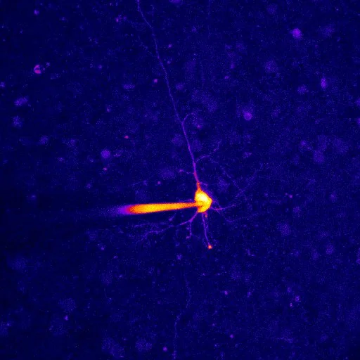
A fiery cell,
Yau Ming Chang, 2022
Calcium imaging · Mouse prefrontal cortex
This image shows pyramidal neurons in layers five and six of a mouse’s prefrontal cortex, recorded using calcium imaging. The technique allows researchers to study changes in calcium within cells. The displayed colours should not, by themselves, be read as a calibrated measure of neuronal activity.
Image: Yau Ming Chang